This project was supported by the Denison Research Scholarship.
I analysed the diffusion dynamics of neural activity using a single subject from the WU-Minn Human Connectome Project. The subject was a young adult (age 22-35) and went four 15-minute scans (1 frame per 0.72 s) in a resting state (relaxed fixation on a bright cross-hair in a darkened room).
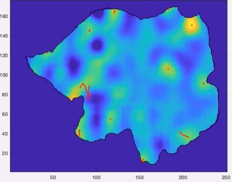
The above figure shows the trajectories of active regions on a 2D map of the subject’s left cerebral cortex hemisphere. This was measured at a single time point, and the warmer regions indicate increased activity.
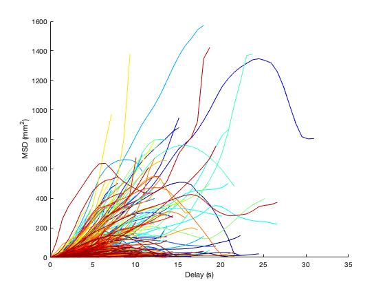
The diffusion analysis was performed using mean square displacements (MSD) of the 874 active trajectories (as shown above). I also computed the diffusion exponent of six regions of interest from the log-log representation of MSD. The regions of interest were chosen based on task-positive regions (Frontal Eye Field, Intraparietal Sulcus, Middle Temporal region) and task-negative regions (Medial Prefrontal Cortex, Posterior Cingulate Cortex, Lateral Parietal Cortex). T-tests were then performed to determine any statistically significant differences between the diffusion exponent of a region of interest and the whole cortex.
A superdiffusive process of the propagation of active regions was found, with the task-negative Posterior Cingulate Cortex displaying faster diffusion processes relative to the active regions in the cerebral cortex. Conversely, the task-positive Frontal Eye Field displayed slower diffusive processes. This was because the default mode network becomes more active during the resting-state that the subject was in, and the task-positive regions are anti-correlated.
Interestingly, the remaining regions of interest had no statistically significant differences in the diffusion exponent compared to the cortex. This could be attributed to the low sample size of trajectories within the regions of interest, which may not adequately represent their neural dynamics.