I optimised a computational model to investigate the electrical response of neuromorphic nanowire networks for retinal interface application. In this model, the nanowire network is simulated as a graph representation, where the nodes corresponds to silver nanowires and the edges represent silver-PVP-silver junctions. Two nodes that are the furthest apart are chosen to be the source and drain nodes, and a voltage bias (e.g. DC stimulus, retinal ganglion stimulus) is applied between them. As voltage is applied, a current path of the least resistance is formed, which is shown by the directed edges. This path is determined by the connectivity of the network, as well as, the Kirchoff’s voltage laws that are solved at each time point.
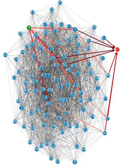
In the above figure, a 100-nanowire, 2285-junctions network is represented, where the green and red nodes are the source and drain, respectively.
I conducted multiple experiments to optimise the parameters for a retinal application of a nanowire network. These parameters included: lengthening the duration of stimulus by repeating the original stimulus a number of times, changing the order of magnitude of voltage levels (e.g. to a biologically relevant voltage in mV), decreasing the set and reset voltages by half from Vset = 0.1, Vreset = 0.01 to Vset = 1.5e−7, Vreset = 1.5e−8, and testing a range of network densities from 100-nanowires and 261-junctions to 700-nanowires and 14533-junctions.
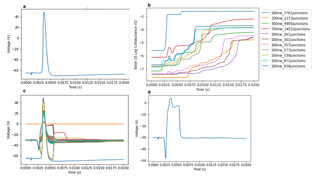
The above figure shows the electrical response of a nanowire network (100 nanowires, 3761 junctions) with a retinal ganglion spike (V). Subfigure a) shows the retinal ganglion spike stimulus. b) shows that increasing network density increased the conductance in response to stimulus. c) shows the diverse range of voltages of all nanowires in response to stimulus, which parallels physiologically-based responses. d) shows the voltages of an individual nanowire in response to stimulus. This voltage profile exhibits a depolarising after-potential (at 5 ms) and subthreshold oscillations (from 10 ms onwards), which are characteristic of a biological neuron.
Hence, the project demonstrated diverse electrical responses produced by neuromorphic nanowire networks. The use of biologically relevant voltages offers strong support for these networks to be researched for future retinal interface application.